Cognitive Science 320
Spring 2011

Synapses and Synaptic Transmission:
Main Concepts of Synaptic
Transmission: Analysis of synaptic transmission and synaptic
potentials requires an understanding of all of the following:
- changing permeability of the membrane to
different ions
- equilibrium potentials of different ions
- passive, electrotonic spread of
subthreshold currents
- mechanisms that are responsible for
production of action potentials
- Soooo, everything you have
learned so far is absolutely necessary for understanding synaptic
processes.
Types of synapses
-
Images of synapses
- Details of chemical
synapses and their neurotransmitters
- Involves
- changing permeabilities of membrane to
different ions
- equilibrium potentials of different ions
- electrotonic (passive) current spread
- mechanisms responsible for production of
the presynaptic action potential
- Presynaptic events -> diffusion through
the synaptic cleft -> postsynaptic events
- presynaptic mechanisms
synapse animation
- regulation of release of
neurotransmitter
- The sequence of events:
- action potential comes into the
axon terminal (voltage-gated Na+ channels and
voltage-gated K+ channels are activated),
depolarizing the presynaptic axon
- depolarization caused by the
action potential activates voltage-gated Ca+2
channels
- Ca+2 moves into the
presynaptic axon terminal through the voltage-gated Ca+2
channels, further depolarizing the axon terminal.
- Ca+2 activates a
series of biochemical reactions which end in the release of the
contents of the synaptic vesicle from the presynaptic axon into
the synaptic cleft.
- The vesicle is somehow pushed to the
'active zone' and the vesicle membrane
fuses with the cell membrane of the presynaptic cell.
- the activation of the vesicle
and fusion with the membrane require many biochemical reactions
between molecules used to mobilize the vesicle from the
cytoskeleton AND biochemical reactions between molecules which
facilitate fusion of the vesicle membrane and cell membrane of
the presynaptic cell. (See Figure 2.30 and 2.32 on pages 48 and
50)
- presynaptic mechanisms
- recycling of vesicle membrane occurs
- this allows maintenance of the
axon terminal's surface area
- this recycles vesicle membrane,
so the cell does not have to make new vesicles each time
transmitter is released.
- Removal of transmitter from the
synaptic cleft
- is necessary to decrease the
length of time that the neurotransmitter can interact with the
postsynaptic receptors
- occurs through one of two main
mechanisms
- (1) reuptake of the intact
neurotransmitter by the presynaptic axon terminals
- Examples: At
the synapses that use the following
neurotransmitters there are reuptake molecules (membrane
proteins) in the membrane of the presynaptic cell, which
quickly and efficiently remove the neurotransmitter from
the synaptic cleft and bring it into the presynaptic cell.
- Examples of neurotransmitters that use
reuptake mechanisms
- norepinephrine (SNRIs
block both norepinephrine reuptake and serotonin reuptake)
- are used as anti-depressants to
increase elevation in mood
- amphetamines affect dopamine
and norepinephrine
- serotonin (SSRIs
block uptake of serotonin: Prozac, Paxil, Celexa)
- are used as anti-depressants to
increase elevation in mood
- used to treat depression,
anxiety and obsessive-compulsive disorder
- dopamine
- creates feelings of euphoria
- reuptake blockers include:
cocaine, methylphenidate, Wellbutrin
- glutamate
- NMDA
receptors
- three types of non-NMDA
receptors
- g-amino
butyric acid (GABA)
- creates sedative, muscle
relaxant effects
- Diazepam blocks reuptake
of GABA
- glycine
- choline (after
acetylcholine is broken down, choline is taken into the
presynaptic nerve terminal for reuse
- (2) enzymatic breakdown of the
neurotransmitter by enzymes on the surface of the
postsynaptic cell
- Example:
At the neuromuscular junction acetylcholine (ACh) is
broken down into acetate and choline by the enzyme
acetylcholinesterase. The choline is then taken up
by the presynaptic nerve through transporter
mechanisms. ACh
----> acetate + choline
(which is reused)
- postsynaptic mechanisms (Figure
of ionotropic and metabotropic receptors)
(Your book illustrates this in Figure 2.29
with few details.)
- (1) quick-acting ion channels
- Produce a quick change in
membrane potential when the neurotransmitter binds onto the
receptor for the neurotransmitter. The receptor contains
an ion channel. When the neurotransmitter binds to the
receptor it activates the channel to open (in most cases it
opens, but in some it can close when the neurotransmitter binds
to it).
-
EPSPs (excitatory postsynaptic
potentials)
- push the cell to threshold for an
action potential
- usually involves allowing positively
charged ions to enter the cell, e.g., Na+ or Ca+2
- glutamate
NMDA receptors
- nicotinic acetylcholine
receptors,
single
channel recordings
-
IPSPs (inhibitory postsynaptic
potentials)
- keep the cell from achieving
threshold for an action potential
- involves adjusting the membrane
potential to a level which will keep the cell from reaching
threshold.
- this can be done by activating
transmitter receptors which increase the permeability to Cl-.
The cell will then hyperpolarize.
- this can be done by activating
transmitter receptors which increase the permeability to K+.
The cell will then hyperpolarize.
- this can be done by closing the
pore of transmitter receptors, thus decreasing the permeability
to Na+. The cell hyperpolarizes because
normally there is a very small leak to Na+.
When this is stopped the cell hyperpolarizes.
-
Think about this: some inhibitory postsynaptic
potentials might depolarize the cell. How could
this be?
-
single channel recordings
from GABAA receptors showing the effect of
pentobarbital on channel openings
-
single channel
recordings from
glycine receptors
- A look at measurement of ion
flow through neurotransmitter-activated single channels
- Two types of presynaptic
voltage-gated Ca2+ channels -
calcium
- (2) sustained acting channels
-
G-protein coupled receptors (from Bear, Connors, Paradiso, p 115)
- neurotransmitter molecules
bind to receptor proteins
- receptor proteins activate
small proteins, called G-proteins, which are free to move
along the intracellular face of the postsynaptic membrane.
- the activated G-Proteins
activate 'effector' proteins.
- See G-protein interactions
and intracellular signaling handout.
- these may alter proteins
(enzymes)
- these may alter permeability of
other ion channels
- these may
alter gene expression
- amplification of the initial
signal: neurotransmitter into activation of multiple
intracellular molecules and pathways
-
cAMP pathways in the cell
-
Review of presynaptic and postsynaptic mechanisms
- Neurotransmitter systems in the
brain (Figure 2.34)
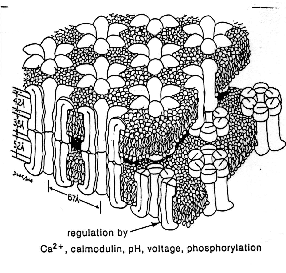
- Details of electrical
synapses
(animation)
- characteristics
- there is no synaptic cleft
- the cytoplasm is continuous through
closable pores
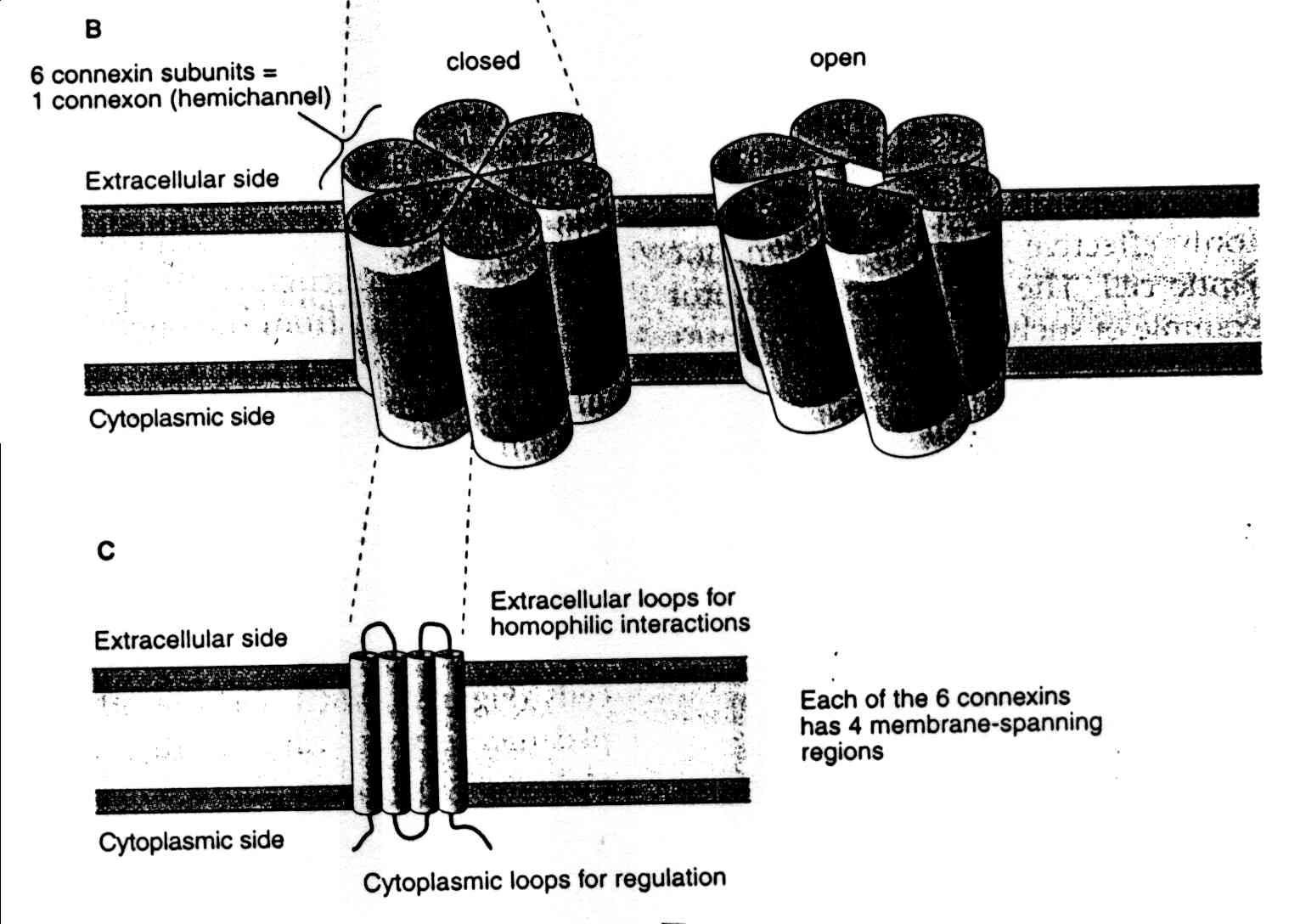
- pores consist of membrane proteins that
form a connexon
- gap junctions (connexin
molecules)
- six connexin molecules form a
hemi-channel (a connexon)
- the principles of electrotonic current
flow are what cause current (information) to pass from cell to cell.
This depends on the length constant of the cells.
- they are faster than chemical synapses
- mechanisms of action
- electrical synapses are used
primarily to send depolarizing signals
- they are used to synchronize a group
of cells
- they can close or open in response
to cellular conditions
- increased intracellular Ca+2
can decrease current through the synapse
- decreased pH in the cell can
decrease current through the synapse
- addition or removal of a phosphate
group can also influence current flow through the pore
- molecules less than 1000 daltons can
pass through the open pore from cell to cell (1 dalton = the weight of
a hydrogen atom)
- Advantages of electrical synapses
- they are faster than chemical synapses
- there is no synaptic delay associated
with release of neurotransmitter
- they allow synchronization of many cells
quickly
- can transmit other cellular signals
(such as chemicals) to influence the connected cells
- Disadvantages of electrical synapses
COMPARED to chemical synapses
- a small presynaptic axon terminal cannot
activate a large postsynaptic cell
- there cannot be amplification of the
synaptic signal
- a presynaptic action potential cannot be
converted into an inhibitory postsynaptic potential (IPSP).
- cannot be modified or modulated to the
same extent that transmitter release from chemical synapses can be
regulated.
-
Example
of electrical synapses in Aplysia.
- Synaptic integration
- EPSPs (excitatory postsynaptic
potentials)
- push the cell to threshold for an
action potential
- IPSPs (inhibitory postsynaptic
potentials)
- keep the cell from achieving
threshold for an action potential
- EPSPs and IPSPs sum within the dendrites
and cell body.
- usually there are no voltage-gated
membrane proteins in the membranes of the dendrites and cell body.
- The axon hillock (action potential
generating site), is usually located where the axon exits the cell body
of the neuron. The electrical signal (sum of EPSPs and IPSPs)
needs to be above threshold when it reaches the axon hillock for the
cell to fire an action potential.
-
The
closer the synapse is to the axon hillock, the stronger the current
from its EPSP or IPSP will be, and the more effect it will have on
whether an action potential is produced or not.
- Spatial
summation: Spatial summation
of EPSPs: when two or more presynaptic inputs are active at the same
time, their individual EPSPs add together.
- Temporal
summation: Temporal summation
of EPSPs: when the same presynaptic fiber fires action potentials in
quick succession, the individual EPSPs add together.
- Timing of synaptic inputs to the
postsynaptic cell as well as the spatial location of synapses are
therefore of critical importance in synaptic integration.
- Crab
stomach pattern generator
- Swimming
pattern generator
Information on Glutamate receptors

